


Professors Hai-Ling Margaret Cheng, Leo Chou, Daniel Franklin, Naomi Matsuura, and Cari Whyne and their collaborators have received a combined funding of $2.6mil+ from the Canadian Institute of Health Research (CIHR) as a part of the 2024 Spring CIHR Project Grant.
The CIHR Project Grant program funds innovative health research projects with the potential to significantly advance health knowledge, healthcare, health systems, and health outcomes.
A complete list of winners can be found here. Read more about the project proposals below.

A Novel Targeted MRI Probe for Safe and Early Detection of Fibrosis
Principal Investigator: Hai-Ling Margaret Cheng
Funding Amount: $875,926
Professor Hai-Ling Cheng’s project aims to improve the detection and treatment of fibrosis, a type of tissue scarring that can occur in vital organs like the heart, brain, lungs, and liver after injuries such as heart attacks, high blood pressure, viral infections, diabetes, or alcohol abuse. Normally, scarring is part of the healing process, but in fibrosis, this process goes out of control, leading to excessive scar tissue and eventually causing organ failure.
Currently, fibrosis is difficult to detect until it is very advanced, at which point treatment options are limited to organ transplantation. This makes it hard to catch the disease early when it might be stopped or reversed, and it also hinders the testing of new anti-fibrosis drugs.
To tackle this problem, the researchers are developing a new imaging agent that can detect fibrosis at early stages. This agent specifically binds to collagen, the main component of scar tissue, and shows up clearly on MRI scans, which are safe and non-invasive. The agent is also naturally eliminated by the body, making it a safe option for patients.
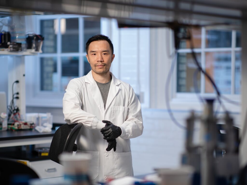
DNA Nanofabrication to Manipulate B cell
Responses
Principal Investigators: Bebhinn Treanor & Leo Chou
Funding Amount: $1,002,150
Professor Leo Chou and his collaborators proposes to develop safer and more effective vaccines using small parts of pathogens, called antigens, and helper molecules known as adjuvants. Instead of using whole pathogens, which can be risky, this approach uses only the necessary components to stimulate an immune response. The findings will help create better vaccines and treatments for diseases involving B cells.
The researchers propose to use synthetic DNA structures to precisely arrange the antigens and adjuvants. These structures allow for exact control over their shape and spacing, mimicking real pathogens. The team will study how these custom designs enhance immune cell activation and antibody production.
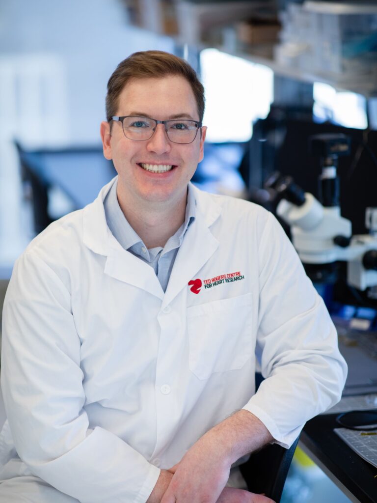
Advancing Pulse Oximetry Accuracy and Equity through Wearable Spectroscopy Devices
Principal Investigator: Daniel Franklin
Funding Amount: $879,750
Professor Daniel Franklin’s project focuses on improving pulse oximeters, devices that measure blood oxygen levels by shining light through a patient’s finger. Pulse oximeters are essential in hospitals, especially for critically ill patients, including those with heart failure, COVID-19, or undergoing surgery.
Recent studies have shown that some pulse oximeters inaccurately read higher blood oxygen levels in patients with dark skin, potentially putting them at risk by making their condition appear safer than it is. To address this issue, the Professor Franklin and his team have developed a new wearable device that uses multiple colours of light to better account for skin tone variations. They have also created an algorithm that adjusts for melanin levels, ensuring more accurate readings for everyone.
The project involves testing and refining this new device and algorithm. These studies will help compare results in both controlled and real-world settings, providing a comprehensive understanding of the bias and potential solutions. The goal is to demonstrate to the medical community and health markets that more equitable pulse oximeters can be designed, leading to fairer healthcare for all Canadians.
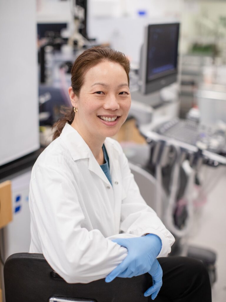
Enhanced Drug Delivery to Lung Cancers using Endobronchial Ultrasound (EBUS)
Principal Investigators: Naomi Matsuura & Kazuhiro Yasufuku
Funding Amount: $753,526
Professor Naomi Matsuura and her collaborators aim to improve lung cancer treatment by making chemotherapy drugs target cancer cells more precisely, reducing harm to healthy cells and minimizing side effects. Lung cancer is a leading cause of death, and current chemotherapy treatments affect both cancerous and healthy cells, causing severe side effects.
In this study, scientists and doctors will use a method involving endobronchial ultrasound (EBUS), a tool commonly used to diagnose lung cancer, and micrometre-scale bubbles, which are safe for imaging blood vessels with ultrasound. EBUS will stimulate these bubbles as they travel through the blood vessels in the tumour, temporarily creating small openings in the tumour’s blood vessels. This allows the chemotherapy drug to flow directly into the lung cancer tumour, sparing normal tissue.
By using this technique, lower doses of the chemotherapy drug can be used, effectively treating the tumour with fewer side effects. The project will test the best conditions for this treatment, including the settings for EBUS, the amount of bubbles, and the timing and concentration of the drug injection. The goal is to find the optimal treatment conditions that can eventually be used to help lung cancer patients.
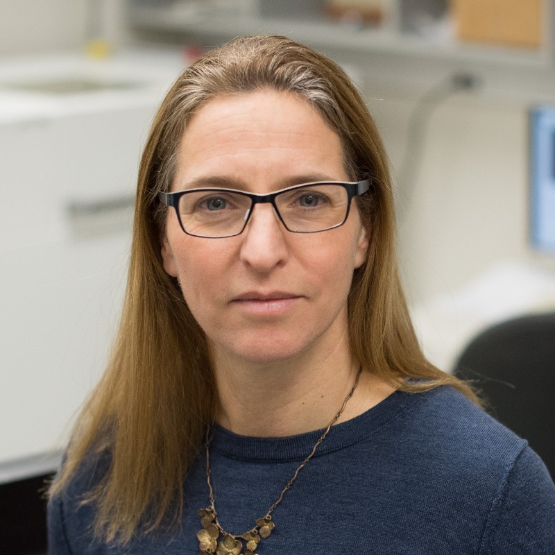
Stereotactic Radiotherapy for Spinal Metastases: Quantifying the Impact on Musculoskeletal Health
Principal Investigators: Cari Whyne; Michael Hardisty; Arjun Sahgal
Funding Amount: $100,000
Dr. Cari Whyne and her collaborators will focus on understanding how a cancer treatment called stereotactic body radiation therapy (SBRT) affects bone and muscle health in patients with cancer that has spread to the spine. SBRT is very effective in treating spinal tumours, but as patients live longer after treatment, it’s important to study its long-term effects on their bones and muscles.
The researchers will analyze a large collection of existing medical images, such as CT and MRI scans, to see how SBRT impacts conditions like osteosarcopenia (loss of both bone and muscle) and spinal alignment over time. Additionally, the study will follow patients for three years after their SBRT treatment, using advanced imaging techniques to measure changes in their bones and muscles. The researchers will also track patients’ pain levels, physical function, strength, and quality of life.
By improving the understanding of how SBRT affects bone and muscle health, this research aims to identify patients who are at high risk of musculoskeletal decline. This will help doctors tailor treatments that not only target spinal tumours but also preserve bone and muscle health, enhancing patients’ overall well-being.