

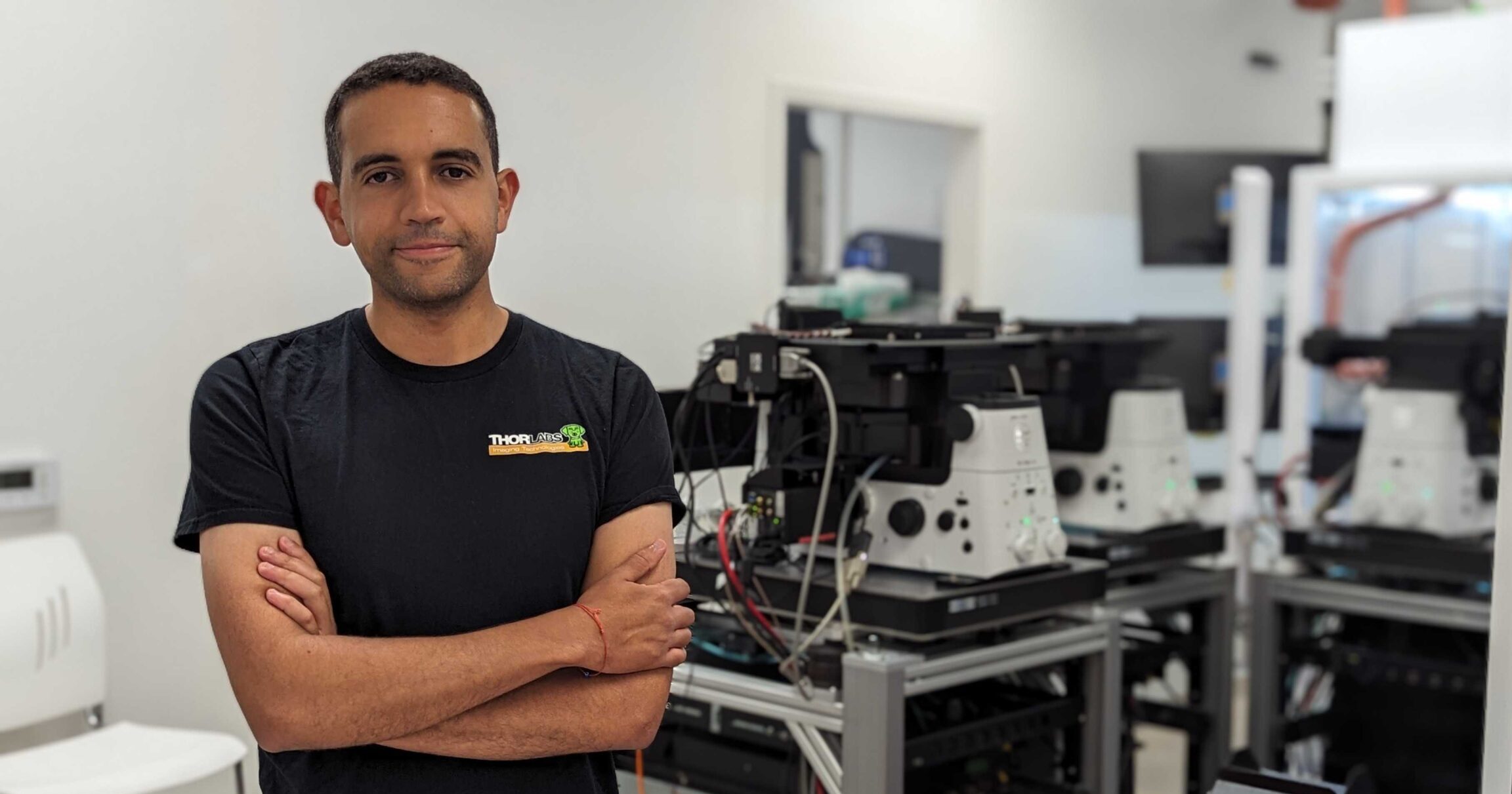
Amine Driouchi (BME2019) embarked on his science journey at King’s College London in biochemistry, and this eventually led to his PhD work in Dr. Chris Yip’s lab at the University of Toronto. Now he is working as a senior scientist for Eikon Therapeutics, a California-based biotechnology company that develops drug discovery technologies using super-resolution microscopy. Here, Amine shares his academic journey and what he is doing now at Eikon.
Can you briefly describe your academic journey before joining Eikon?
Before joining Eikon Therapeutics, my academic journey was driven by a passion for understanding biology at its fundamental level. During my undergraduate studies in biochemistry at King’s College London, I realized the need to move beyond descriptive approaches and delve into single molecule biophysics to explore molecular complexities.
I pursued labs in biophysics during graduate school and joined Chris Yip’s lab at the University of Toronto (U of T). Chris’s mentorship encouraged curiosity and fearlessness, allowing me to take on challenging and risky projects.
After U of T, I spent two years in Chicago, furthering my personal growth in a research environment. Throughout my journey, I experienced various international moves, which shaped my ability to manage stress and adapt to new environments.
The desire to apply single molecule tracking and super-resolution techniques to drug discovery motivated me to build my own platform before discovering Eikon. Connecting with Eikon’s team, I realized it was the perfect fit for my mission. After joining Eikon, I became part of an incredible journey with a small team, pushing our mission forward. The experience felt like years of work with brilliant minds tackling fascinating scientific questions.
My driving force has always been the underlying scientific questions and the desire to make methods that truly help us understand biology. Success in drug discovery is essential, but even if we do not achieve it, our efforts to advance scientific knowledge are valuable and fulfilling.
As a senior scientist, what is your day to day like?
As a senior scientist at Eikon, my day-to-day work has evolved significantly since I joined the company when it was still relatively small with around thirty people. Back then, my role was quite diverse, and I worked at the interface of biology and engineering, utilizing my expertise in advanced microscopy, particularly super-resolution microscopy, which I had gained during my PhD and postdoc.
During the early days, close collaboration with various departments and team members was essential as we collected vast amounts of data from numerous well plates containing thousands of cells and millions of trajectories. Our primary focus was on developing the platform, ensuring we had the necessary hardware and software to effectively process and interpret the data. Additionally, we worked closely with the automation team to identify and resolve any issues in the system, making our assays more robust and dependable for day-to-day operations.
As Eikon experienced rapid growth and expanded to almost four hundred employees, roles and responsibilities evolved. Now, individuals like me are encouraged to specialize further. Currently, I am deeply involved in early research and development, collaborating with colleagues who share a similar background. Together, and along with my team, our task is to explore new possibilities and determine the platform’s strategic direction for the next two to three years.
The journey at Eikon has been incredibly dynamic, witnessing the company’s remarkable growth and adapting my role accordingly. From overseeing a broad range of tasks in the initial stages to now increasingly focusing on specialized research and development efforts, I continue to find great excitement and fulfillment in contributing to Eikon’s innovative projects.
What does Eikon specialize in?
At Eikon Therapeutics, our specialization lies in examining protein motion in living cells using our innovative technology platform. We focus on gaining valuable insights into drug target engagement, protein disruptions, and subpopulations within cells. Our approach involves single particle tracking, which allows us to move beyond traditional methods and capture the full spectrum of protein diffusion speeds under different perturbations. This comprehensive understanding of target behavior and interactions with cofactors over time sets us apart.
We are also expanding our capabilities beyond single particle tracking. We have a range of advanced microscopy techniques at our disposal, and we continuously strive to improve and explore new measurements with our high-end cameras and optical components. We have also invested in building orthogonal capabilities, including biochemistry, chemistry, and assay biology groups, enabling us to manage protein purification, run luciferase, immunofluorescence assays, and even conduct medicinal chemistry internally.
Having medicinal chemistry capabilities has been a notable change for us. It allows us to rapidly iterate on molecule modifications based on data from various assays, leading to faster progress and innovation.
What is the advantage of using microscopy to examine biological processes inside the cell, in real time?
Using microscopy to examine biological processes inside the cell in real time offers us a significant advantage in our work. It allows us to tackle the complexity of these processes by delving into the intricacies and variations within individual cells, rather than averaging out data as in traditional biochemical methods like western blots.
This detailed information gives us valuable insights into how several factors regulate cells, their spatial distribution, and the heterogeneity brought about by intracellular components. With this level of understanding, we aspire to specifically target subsets of proteins or cells that are relevant to a particular therapeutic indication. This targeted approach can give us a competitive edge in our drug discovery efforts.
In our field, there is a tendency to average granular datasets for simplicity, but we recognize the importance of questioning the origins of trends. By delving deeper into the details, we can improve drug selectivity and specificity, leading to potentially cleaner drugs. As our research advances, we have progressed to more complex and costly systems, including animal and clinical trials, and having a better understanding of the mechanisms and biology in these systems aids our medicinal chemistry efforts.
Can you give me an example of a use case, where using these advanced microscopy techniques can help solve a biological problem?
Using single particle tracking, we can infer whether a protein exists as a free monomer, a heterodimer, or part of a larger complex based on diffusion coefficient measurements. This provides us with a deeper understanding of how proteins associate with their cofactors. At Eikon, we thrive on operating outside the norm and exploring uncharted territories. By focusing on promising areas that others might overlook, we gain a competitive edge in drug discovery and target identification.
Our platform’s emphasis on protein-protein interactions and understanding their behavior over time is a notable change. This level of insight is rarely achieved in living cells through conventional approaches, and it holds immense potential for refining our understanding of cellular function and protein dynamics.
In fact, we are currently engaged in exciting collaborations that leverage our platform’s scale and capabilities to push the boundaries of research. Together, we are making significant strides in studying protein dynamics as they transition from monomers to higher-order oligomers under specific conditions.
What is a major challenge for you and the company right now?
In my view, the evolution of super-resolution technology in the next few years will depend on how we tackle the challenges currently faced in the field of microscopy, especially in super resolution microscopy and single particle tracking. One major challenge is the proliferation of methods and papers with questionable quality and contributions, which can lead to distractions and dilute research efforts.
However, I have noticed that researchers tend to rise to the occasion when faced with challenging problems, and they actively start experimenting and innovating. To move forward, it is crucial for us to refocus our efforts on addressing the most critical biological questions using super resolution microscopy. This involves customizing imaging techniques to suit the specific biology of interest, effectively bringing the microscope to the biology rather than forcing the biology to fit the microscope.
At Eikon, I encounter a similar challenge in a growing company, where some folks may perceive the technology as fixed and inflexible. However, I advocate for maintaining a sense of flexibility and customization, as this allows us to address a wide array of biological questions, considering the diverse range of targets and cellular processes.
In the context of super resolution microscopy, the role of hardware and software is paramount. For instance, properly studying protein motion in cells is a non-trivial task, especially when dealing with a high labeling density, where reconnection of individual detections become particularly challenging. To ensure the reliability and significance of our assay results, we need trustworthy outputs and downstream analyses that provide a genuine assay window.
In conclusion, the key to the evolution of super-resolution technology in the coming years lies in embracing change and refocusing our efforts on addressing biologically relevant questions while adapting imaging approaches to suit the specific biological context, whether in a research lab or a pharmaceutical company. By doing so, we can make significant strides in advancing super-resolution microscopy and its applications in understanding complex biological processes at a finer scale.