

In a recent study, researchers from the University of Toronto employed a unique state-of-the-art imaging technique for deep tissue imaging, that has enabled the monitoring of peri-implant bony healing biology in action. This technology can lead to a better understanding of the healing process, allowing researchers to leverage this knowledge to develop faster therapeutic approaches with the use of biomaterials for the future.
This finding was recently published in the journal Biomaterials, as a result of collaborations between state-of-the-art imaging facility at the University Health Network (UHN), leading providers of dental implants, and the Toronto-based mesenchymal stem cell company, Tissue Regeneration Therapeutics Inc.
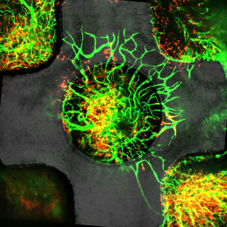
During bone healing, either from injury or from an implant placement, specialized cells called mesenchymal progenitors are recruited by the body to the site of injury to facilitate healing. The mechanisms and dynamics underlying the migration and recruitment of these critically important progenitor cells into the wound site have, until now, remained a mystery.
“Our results took our understanding of peri-implant healing to a whole new level as it allowed us to track the provenance and dynamics of mesenchymal cells in real-time at a single-cell resolution.” Said Dr. Niloufar Khosravi, the lead author of this study.
Being able to visualize and monitor the cells at a high resolution had been previously difficult, but the authors were able to employ a new imaging modality to monitor the cell recruitment process and discovered new processes that were not known before. This technique was developed in collaboration with scientist at DaCosta lab, Princess Margaret Cancer Center.
“We were also able to show that these cells differentiated to repair both the bone tissue and also control the newly formed vasculature, a finding that was uniquely the result of our imaging techniques, and which has not been previously reported in the literature.” Said Khosravi.
Although the research findings were focused on the context of dental healing, the authors believed the knowledge can be applied to the healing of other tissue types.
“We hypothesized that the proliferative bloom of mesenchymal precursors, which inevitably had a profound effect on the local the wound healing environment, could be instrumental in driving the transition from an acute inflammatory to a reparative tissue response.” Said Dr. Davies, “Such deepening of our understanding of wound healing biology, by real-time imaging, provides new insights into personalized and directed therapeutic approaches.”