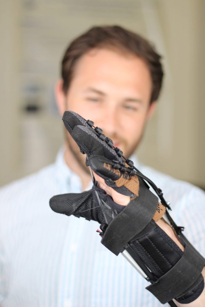
HERO Gloves
Aaron Yurkewich, Illya Kozak, Andrei Ivanovic, Mihailidis Lab
The HERO Glove assists stroke and spinal cord injury survivors to extend their hand and grasp with more force. The HERO Glove enables people to perform activities of daily living like opening water bottles, cutting food and writing independently at home.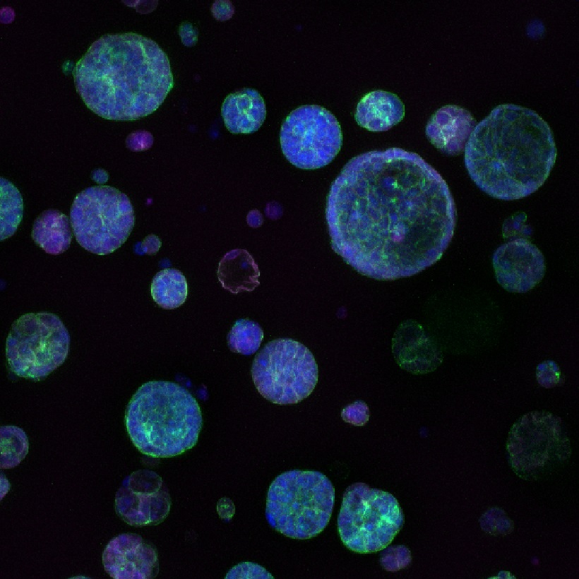
Biomaterials for better cancer therapies
Yihe Wang, Kumacheva Lab | Alexander Baker, PhD, Shoichet Lab
Human luminal breast epithelial cancer cells are grown as multi-cellular spheroids in a microfluidics platform. Blue represents the cell nucleus, green the cell cytoskeleton and red the cell tight junctions, which is a hallmark of 3D culture.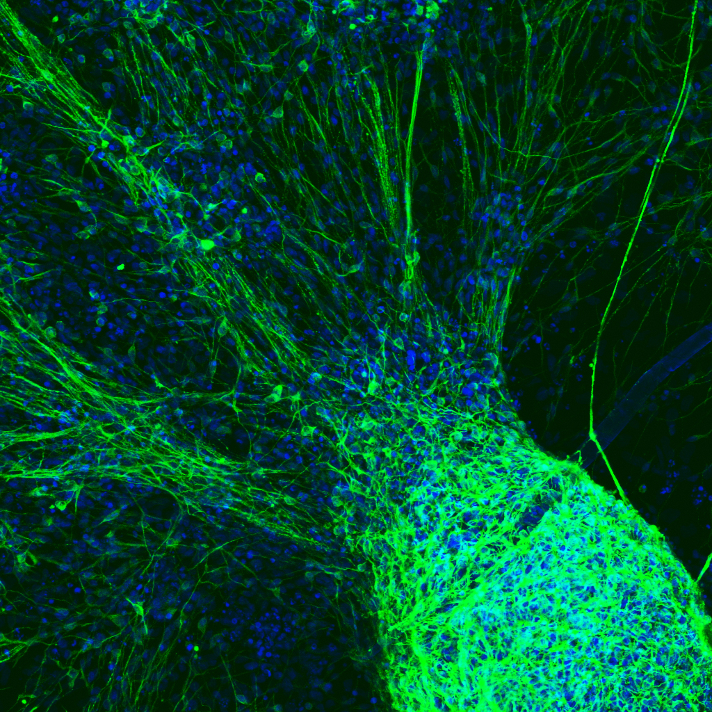
Striving to repair spinal cord damage
Tobias Fuehrmann, Post Doctoral Fellow, Shoichet Lab
Stem cells can be programmed to generate specific cells types. Here cells are differentiated into nerve cells (green) which extend large processes, called axons. The nuclei of all cells are blue.
Simulator for self-driving vehicles
Shabnam Haghzare, KITE Toronto Rehabilitation Institute, Mihailidis lab
At the Toronto Rehab’s driving simulator (DriverLab) located at the Toronto Rehabilitation Institute can look at the safety and acceptability among older adults with potential cognitive impairments such as dementia.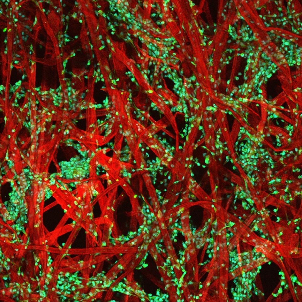
Cancer cells on paper
Simon Latour, Mcguigan Lab
The picture represent cancer cells growing in a paper based 3D environment.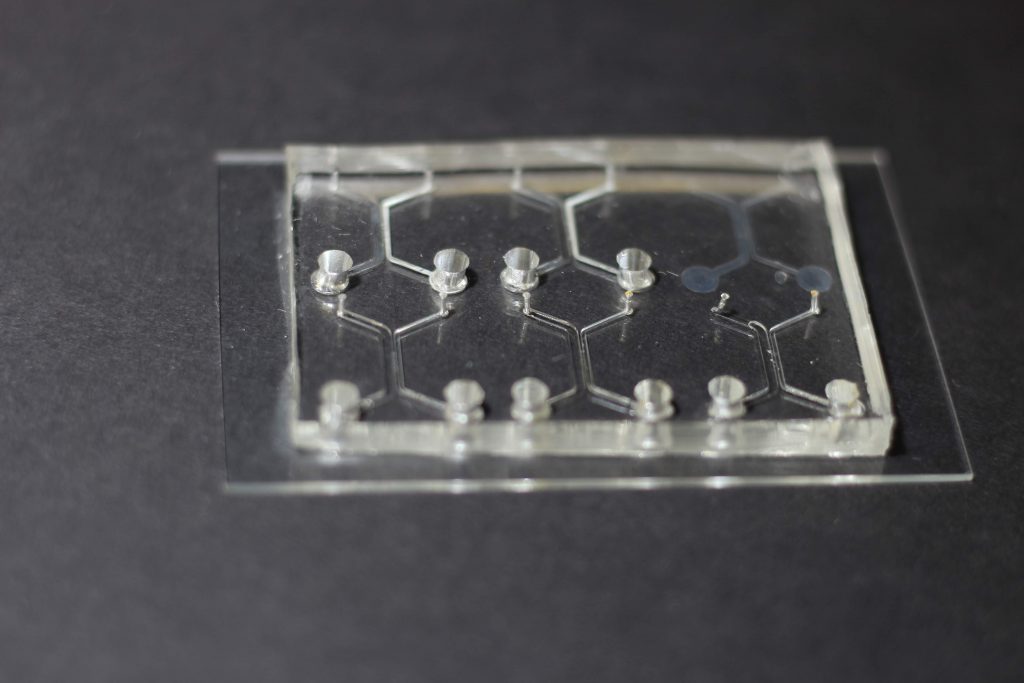
Microfluidic device for modeling cancer metastasis
Christina Mei, Kevin Middleton, You lab
This is a microfluidic cancer extravasation tissue platform that integrates stimulatory bone fluid flow and real-time bi-directional signaling between multiple cell populations, as to investigate the role of osteocytes in the mechanical regulation of breast cancer bone metastasis.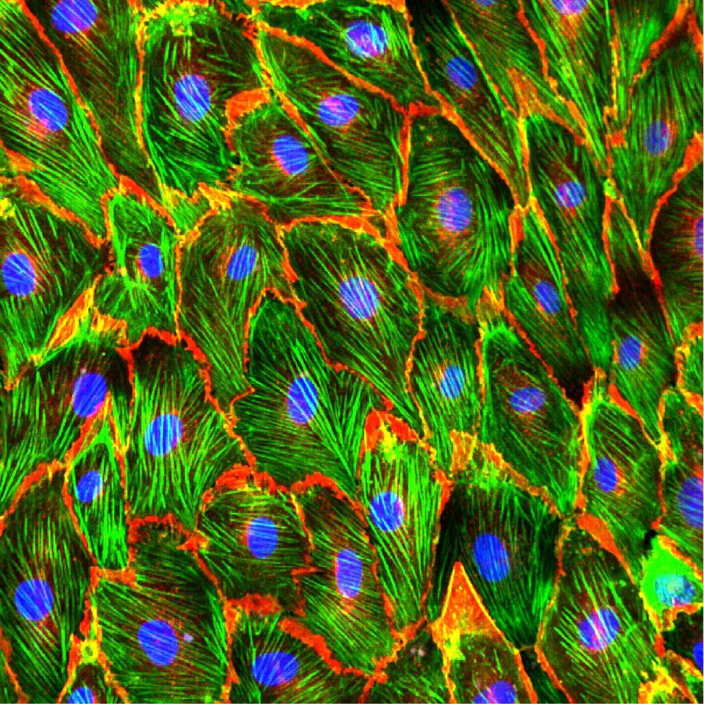
Modeling the blood vessels
Yih Yang Chen, Chan Lab
Human Umbilical Vein Endothelial Cells (nuclei stained in blue) are grown within a microfluidic channel and subjected to flow shear in order to align their actin fibres (green) in the direction of flow. VE-Cadherin protein expression (red) shows the cell membranes are cross-linked to each other, allowing all of the individual cells to resist being washed away.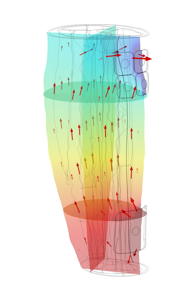
Neural activation of motor nerves
Silviu Agotici, Yoo Lab & Masani Lab
The image shows the finite element (FE) solution for the electric field (heat map) and the current flow (arrows) resulting from the simulation of transcutaneous electrical stimulation (TES) in a 28 cm section of the lower leg (just below the knee to just above the ankle).
These simulation allow us to gain a deeper understanding of the effects of transcutaneous electrical stimulation variables such as electrode shape and positioning, and stimulation magnitude on neural activation.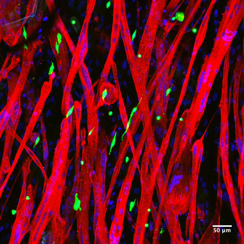
Skeletal muscles
Bella Xu, Gilbert & Mcguigan lab
The mouse skeletal muscle stem cells (green) associate with human skeletal muscle fibres (red) 24 hours after the incorporation of GFP muscle stem cells in 3D human muscle tissue (MEndR).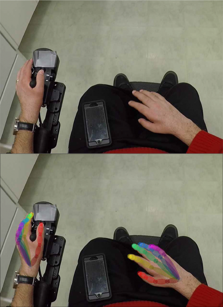
Rehab using augmented reality
Mehdy Dousty, Zariffa Lab
Zariffa and colleagues are applying computer vision algorithms on videos captured from wearable camera to monitor patient hand recovery after spinal cord injury. This can provide more effective clinical evaluations of new interventions through precise outcome measurements.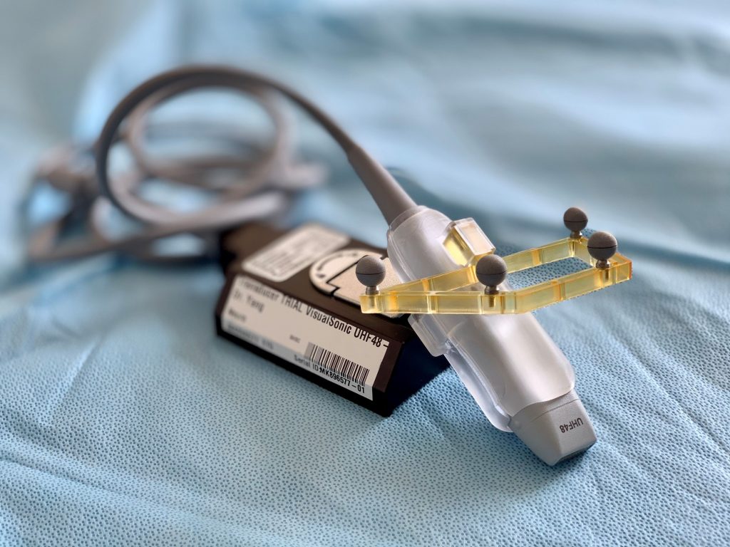
Vascular detection in neurosurgery
Shaurya Gupta, Yee and Yang Lab
This is a 3D printed devices that surgeons use on clinical cases in the Operating Room. This device allows for accurate tracking of the ultrasound probe (and by extension – ultrasound scans) in 3D space and in relation to the patient’s preoperative CT/MRI scans.


