Tissue regeneration has tremendous potential to heal damaged tissues and regrow lost limbs. In the past two decades, researchers have proposed transplanting stem cells or non-cellular scaffolds into the injury site to facilitate tissue regeneration. Stem cells can differentiate into specialized cell types in the body, and the non-cellular components provide growth factors and other nutrients that facilitate tissue regrowth. It is relatively easy to observe tissue regeneration on the skin; however, regeneration of internal organs is more difficult to visualize. Currently, no technology exists that allow researchers or clinicians to monitor non-invasively the tissue regeneration process deep inside the human body.
It is a tremendous challenge to monitor whether these therapies are working. As a patient, the last thing you want is to have these cells implanted and then have a highly invasive surgery a couple of months down the line to check whether the cells are still there,” says Dr. Hai-Ling Margaret Cheng, an IBBME/ECE principal investigator at the Ted Rogers Centre for Heart Research, “We want to ensure that the patient monitoring process is as painless as possible, and one way to do that is to use non-invasive imaging to track the cells and non-cellular components we introduce into the body.”

One of the many projects in Dr. Hai-Ling Margaret Cheng’s lab is to develop track-ers that identify the location of these exogenous components. Coming from a background in medical biophysics and electrical engineering, Dr. Cheng has spent more than 14 years developing technologies that can enhance Magnetic Resonance Imaging (MRI) of the human body. Prior to MRI session, patients may need to intake a contrast agent to improve detection sensitivity and greater signal difference on MRI. However, if we directly incorporate these contrast agents to the stem cells or non-cellular scaffolds, contrast agents can act like miniature trackers in addition to enhancing contrast. In other words, they act as a long-term surrogate in the body. Safety is a foremost concern when it comes to designing contrast agents. While normal contrast agents last in the body for only a few hours before being excreted, contrast agents designed to monitor regenerative medicine components might stay in the body up to a few months. During that time, researchers have to evaluate their interactions with various organs such as the kidney, the blood, and the liver.

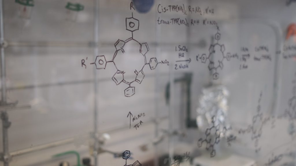
“Traditional contrast agents are completely foreign to the body, and we’ve seen them accumulate in the bone and the brain, leading to toxic effects. So in the context of regenerative medicine we have to design everything from scratch,” says Daniel A. Szulc, a senior member in the Cheng lab who specializes in MRI contrast agent design, “These agents will be traveling through the blood stream, bypassing certain organs and getting into the cells. There’s a design consideration for each step of the way.”
When people get breast implants, they certainly do not want them to be moving around too much, let alone degrade in their body. Our contrast agents could be incorporated in these implants so individuals with these cosmetic surgeries could have their checkups with a non-invasive method like MRI,” says Margaret. “We are planning to bring our tracking technologies into the clinic in the next five years. I think it will be a game changer for everyone who’s involved in tissue regeneration and regenerative medicine”


